

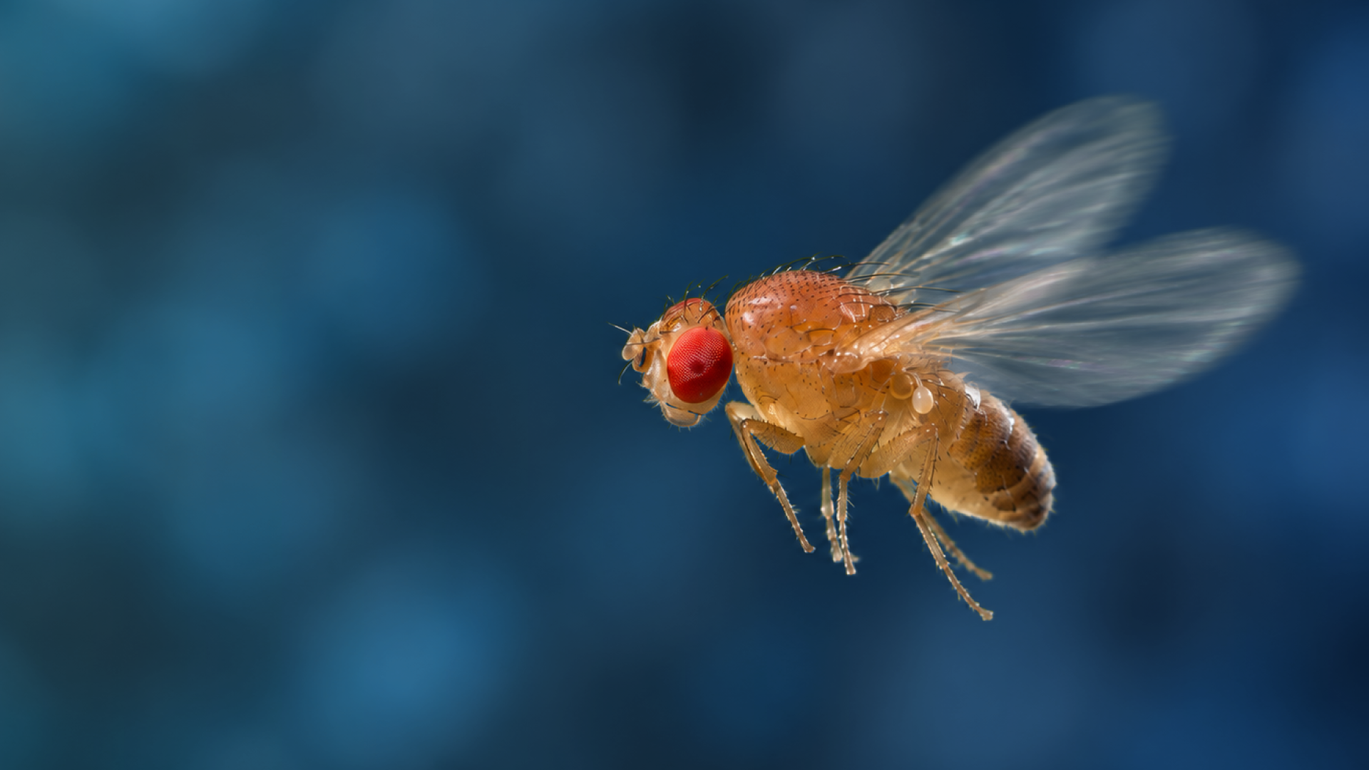
Drosophila Imaging
Drosophila imaging with FEMTO3D Atlas enables high-speed, submicron-resolution monitoring of neural activity to study synaptic integration, sensory coding, and plasticity in precisely defined circuits.
About Drosophila Imaging
Drosophila provide an ideal system for dissecting neural circuits with genetic precision. The FEMTO3D Atlas enables rapid voltage and calcium imaging with submicron spatial resolution, while advanced scanning strategies such as line and ribbon scanning allow detailed interrogation of dendrites, spines, and small neuronal populations. This makes it possible to uncover fundamental principles of synaptic integration, sensory coding, and plasticity at high temporal resolution.
Drosophila Imaging samples


Voltage Imaging

Mushroom body in 3D

FEMTOSmart Resonant scanning

Anatomy Z-stack
Advance your research with Drosophila Imaging
Talk to an application specialist about your research, or book a demo at one of our facilities.