

Voltage imaging
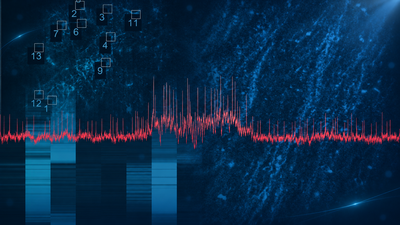
Resolve individual action potentials in vivo, including spikes within fast bursts, at the temporal resolution voltage signals demand.
About Voltage Imaging
Genetically encoded voltage indicators (ASAP, JEDI2P, etc.) have recently attained a level of quality that renders them valuable tools for studying membrane potentials throughout cells. However, a significant challenge has persisted: the lack of sufficiently fast equipment to capture their dynamics. Until now, there has been a shortage of systems capable of imaging at speeds exceeding kHz, approaching the electrophysiological standard of 10 kHz. The FEMTO3D Atlas addresses this gap by enabling high-speed, three-dimensional voltage imaging. Equipped with the real-time motion correction module - the 3D Real-Time Motion Correction - it achieves rapid imaging without motion artifacts and ensures high signal-to-noise ratio (SNR) for precise voltage imaging.
Key features of Voltage Imaging
Indicators

In Vivo Voltage Imaging
In Vitro Voltage Imaging
Single spikes, captured in vivo


In vitro JEDI-2P measurement combined with uncaging

In vivo drosophila imaging

3D voltage recording
Research powered by Femtonics systems


The methods, explained by the people who use them
interview
An interview with Tristan Geiller: Shining a new light on memory formation
webinar
Tune in to the brain - 3D acousto-optic technology in multiphoton microscopy
interview
Interview with neuroscience researcher, Christopher Makinson, PHD.

Every Atlas ships with the team behind it
Installation, training, and ongoing support are handled directly by Femtonics engineers, not a distributor. From first light to advanced experiments, the people who built the system are the ones helping you run it.
Advance your research with Voltage Imaging
Talk to an application specialist about your research, or book a demo at one of our facilities.