

Zebrafish Imaging
Zebrafish imaging with FEMTO3D Atlas enables deep, in vivo volumetric recording of neural structure and activity to study circuit development, reorganization, and behavior over time.
About Zebrafish Imaging
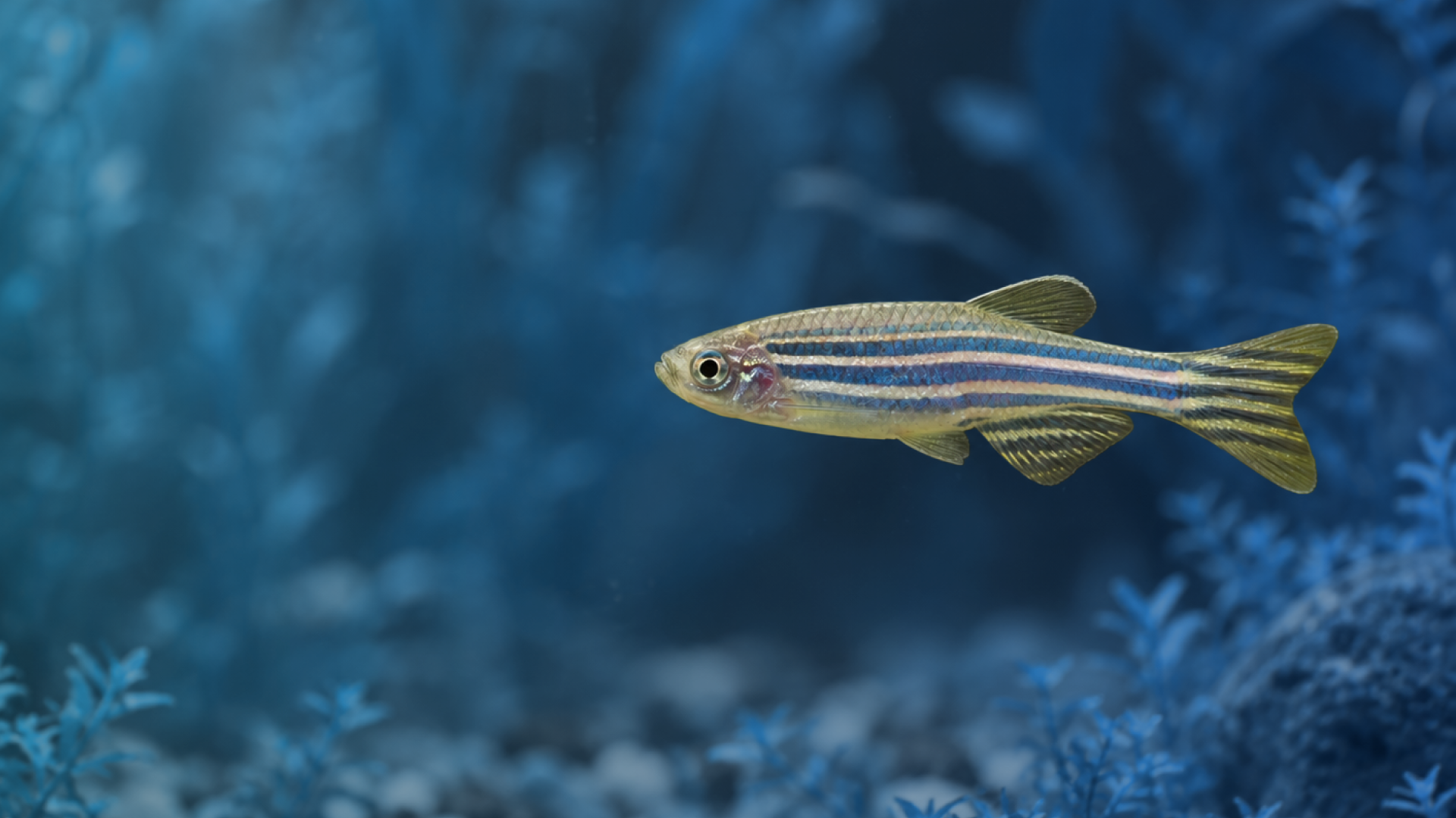
Zebrafish are highly suitable for developmental and systems neuroscience due to their transparency and genetic tractability. The FEMTO3D Atlas supports deep, in vivo imaging across developmental stages, enabling time-lapse and volumetric recordings of neural activity. By capturing both anatomical structure and functional dynamics in intact animals, researchers can study how circuits form, reorganize, and generate behavior over time.
Zebrafish Imaging samples


Timelapse imaging of embryonic zebra danio
Advance your research with Zebrafish Imaging
Talk to an application specialist about your research, or book a demo at one of our facilities.